Skeletal system
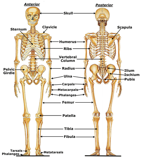
The skeletal system acts as a foundation for the human body. This system includes all of the bones and joints that allow movements and support for the structure of a human. There are many functions that bones can serve, such as producing blood cells in bone marrow. Bones also provide an insertion point for muscles that lie superficial to bones. There are approximately 206 bones in the human body.
The skeletal system is split up into two regions: axial and appendicular. The appendicular skeleton refers to the appendages (arms and legs) and any bones in those areas, whereas the axial refers to every other bone in the body.
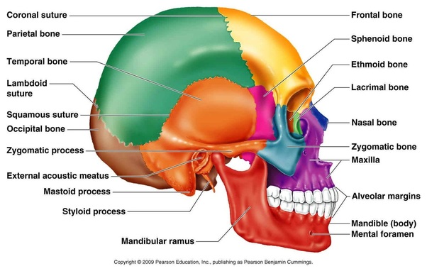
The Skull: Axial
The skull is split into cranial and facial bones. There are a total of 22 bones in the skull, and they serve a unity function of brain protection as well as other soft tissues around the area. During childhood, the bones are somewhat separate allowing room for the brain to grow.
Cranial Bones (8): Some of the bones are in multiples due to the fact that they lie on either side of the head.
Facial Bones (14): Similar to the cranial bones, some of the facial bones are on either side of the head, making them doubles.
The skull is split into cranial and facial bones. There are a total of 22 bones in the skull, and they serve a unity function of brain protection as well as other soft tissues around the area. During childhood, the bones are somewhat separate allowing room for the brain to grow.
Cranial Bones (8): Some of the bones are in multiples due to the fact that they lie on either side of the head.
- 1 Frontal Bone
- 2 Parietal Bones
- 2 Temporal Bones
- 1 Occipital Bone
- 1 Ethmoid Bone
- 1 Sphenoid Bone
Facial Bones (14): Similar to the cranial bones, some of the facial bones are on either side of the head, making them doubles.
- 1 Mandible Bone
- 2 Maxillae Bones (s. Maxilla)
- 1 Vomer Bone
- 2 Palatine Bones
- 2 Nasal Bones
- 2 Zygomatic Bones
- 2 Nasal Conchae (s. Concha)
- 2 Lacrimal Bones
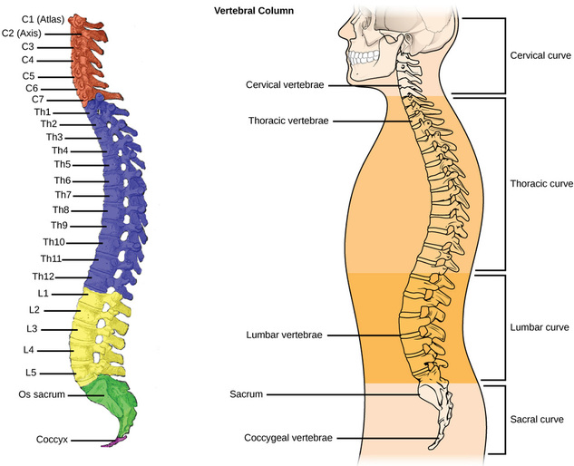
The Spine (Vertebral Column): Axial
The spine plays one of the most important roles in the human body. It plays that of protection, similar to many bones and other structures, but without this support there would be too many disabilities to count. It supports all of the weight of the upper body, causing paralysis if damaged, as well as protecting the spinal cord and many other nerves that control throughout the body. The spine also allows a wide range of movement and flexibility, but if extended past what is allowed, there is severe consequences.
There are 5 main regions of the spine, and they are as follows:
- Cervical - This section is convex and the most superior consisting of seven different vertebra. The seven vertebrae are named C1-C7, based upon the first letter of the name of the region and where they lie superior to inferior. There are two special vertebrae in the cervical curve; C1 and C2. Vertebra C1 is also known as "Atlas," after the Greek titan that held the Earth on his shoulders. The skull moves up and down due to atlas. Vertebra C2 is also known as "Axis," because it allows the skull, and atlas, movement to the left and right.
- Thoracic - There are twelve vertebrae that compose the thoracic region lie within the posterior portion of the chest cavity. These vertebrae are named T1-T12. They are larger and stronger than cervical vertebrae, although much less flexible. The unique function of the thoracic vertebrae is to attach the ribs and support the rib cage that protects the organs within the chest cavity.
- Lumbar - The lumbar curve composes the lower back region. These five vertebrae are named L1-L5. They are larger and stronger compared to thoracic vertebrae, but more flexible too due to lack of rib attachments. This region does a majority of the upper body weight bearing due to their strength, but this is where the origin of back problems reside.
- Sacral - The sacral region is only composed of the sacrum bone which lies between the hip bones. During adolescence, the sacrum begins as five vertebral bones, but fuses together over time in order to provide more adequate support.
- Coccygeal - Normally consisting of 4 vertebrae (Cx1-Cx4) the coccyx performs a simple, yet important function of supporting body weight when sitting down. This section can consist of 3, or 5, vertebrae although the function does not alter. It is commonly referred to as the tailbone; it is homologous to the tailbone in animals.
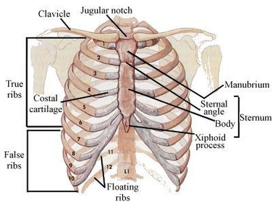
Thoracic Cavity/Rib Cage: Axial
Sternum: There are three parts of the sternum; the Manubrium, the body, and the Xiphoid process (named from superior to inferior). Collectively these bones serve as an attachment place for the ribs/rib cage. The sternum also assists in protecting the thoracic organs such as the heart and lungs.
Ribs/Rib Cage: There are three types of ribs that compose the rib cage; true, false, and floating ribs.
- True Ribs - Pairs 1-7; these ribs directly attach to the spinal column and sternum by costal cartilage.
- False Ribs - Pairs 8-12; these ribs do not have a direct attachment to the sternum, but instead they attach to the cartilage of the ribs above.
- Floating Ribs - Ribs 11 & 12; these ribs do not attach to the sternum at all; only to the spinal column.
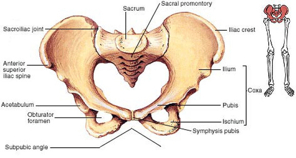
Pelvic Girdle: Axial
The pelvic girdle only consists of three bones; the illium, the ischium, and the pubis. This region also consists of parts of the sacral and coccygeal vertebra.
The pelvic girdle only consists of three bones; the illium, the ischium, and the pubis. This region also consists of parts of the sacral and coccygeal vertebra.
- Illium - The largest of the three, the illium is a large, cup-shaped bone with large crests anteriorly and posteriorly. This bone serves mostly for mobility of the lower extremities, but there are also spines that allow attachment for ligaments and muscles.
- Ischium - This posterior and inferior bone composes ones' rear end, and it supports body weight when one sits down. Many major blood vessels and arteries run through the pelvic region, especially the ischium, allowing blood flow to the legs and feet.
- Pubis - The intermediate bone of the region allows to attachment of pubic bones and is fused together by the pubic synthesis. There is a distinct difference between male and female around this region.
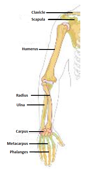
Upper Extremity: Appendicular
The upper extremity consists of the shoulder, arm, hand, wrist and fingers. There 32 bones the correlate with the upper extremity; 2 for the shoulder, 3 for the arm, 8 for the wrist, 5 for the hand, and 14 for the fingers.
The upper extremity consists of the shoulder, arm, hand, wrist and fingers. There 32 bones the correlate with the upper extremity; 2 for the shoulder, 3 for the arm, 8 for the wrist, 5 for the hand, and 14 for the fingers.
The shoulder consists of the clavicle and scapula.
The arm consists of the humerus, radius, and ulna.
The wrist consists of the 8 carpal bones: scaphoid, lunate, triquetrum, pisiform, trapezium, trapezoid, capitate, and hamate (named from lateral to medial & proximal to distal).
The hand consists of metacarpals.
The fingers consists of phalanges.
- Clavicle - commonly known as the "collarbone," serves to connect the sternum and scapula. The clavicle is one of the most commonly broken bones in the body and is used as a landmark due to its easily identifiable location.
- Scapula - commonly known as the "shoulder blade," serves as an attachment for muscles of the arm and shoulder area. There is much muscles padding the bone, therefore it requires much external force to break it.
The arm consists of the humerus, radius, and ulna.
- Humerus - serves a major purpose as an attachment for many muscles on the upper arm, the forearm, and the shoulder. The humerus is the largest bone and only bone in the upper arm. Without the humerus, activities such as writing, throwing, and lifting would not be possible.
- Radius - the slightly shorter bone of the forearm and corresponds with the thumb, or lateral aspect of the arm. The rotation at the wrist is caused by the many muscles that attach at the radius. These muscles also provide much movement for the upper arm and the movements it makes as well.
- Ulna - the longer, larger, and more medial (corresponding to the "pinky" finger) of the forearm bones and many muscles attach here in order to perform movements of the arm, hand, and wrist. The proximal end of the ulna creates the elbow, and when the ulnar nerve at that ulna head is pinched there is a sharp pain that is commonly referred to as "hitting your funny bone," therefore the ulna is the funny bone.
The wrist consists of the 8 carpal bones: scaphoid, lunate, triquetrum, pisiform, trapezium, trapezoid, capitate, and hamate (named from lateral to medial & proximal to distal).
- The carpal bones are tiny bones that are commonly broken, although they can support a lot of weight when needed. These bones compose the wrist and allow for quick support when falling.
The hand consists of metacarpals.
- The metacarpals form the hand, and are also named from lateral to medial. They run from the carpal bones of the wriist to the proximal end of each digit. The thicker, palm layer of the hand is protected by connective tissue, but the other side they can be seen through the skin. The heads of the metacarpals form what are commonly known as knuckles. There are also hand muscles that connect to these bones and allow for function within the remaining parts of the hand.
The fingers consists of phalanges.
- There are a total of 14 phalangeal bones on each hand. They are usually named from lateral to medial, by roman numerals, or more commonly referred to from thumb to little finger. The thumb only has two phalanges due to its size, but the remaining fingers have three phalangeal bones. These bones allow movement and flexibility, such as grasping, for the hand. The hand only serves as a connector piece, but the fingers serve the true function.
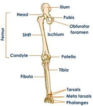
Lower Extremity: Appendicular
The lower extremity consists of the thigh, shin/calf, ankle, foot, and toes. There are only 29 bones that correlate with the lower extremity; 1 for the thigh, 2 for the lower leg, 7 for the ankle,
The lower extremity consists of the thigh, shin/calf, ankle, foot, and toes. There are only 29 bones that correlate with the lower extremity; 1 for the thigh, 2 for the lower leg, 7 for the ankle,
The thigh consists of the femur.
The shin consists of the tibia and the fibula.
The ankle consists of the tarsals.
The foot consists of the metatarsals.
The toes consists of phalanges, similarly to the fingers.
Information for this page is from http://www.innerbody.com/image/skelfov.html
- The femur is the longest, strongest, heaviest bone in the whole human body. All of the body's weight is supported by the femurs during major activities such as walking, running, standing, etc. The hip and thigh muscles, such as the quadriceps femoris, the hamstrings, and the adductor groups also help with much resistance against high tension forces.
The shin consists of the tibia and the fibula.
- The tibia is the stronger, weight-bearing bone of the lower leg that lies medial to the fibula. It forms the knee joint with the femur and the ankle joint with the fibula and tarus. Many muscles anchor onto the tibia in order to perform high tension forces, similar to the femur although not the same. With a fracture of the tibia, the leg will not be able to support and important movements. It will need be immobilized and set for recovery.
- The fibula is the weak, non-weight bearing bone of the lower leg that lies on the lateral aspect. The main purpose of the fibula is to support the ankle joint and the muscles of the lower leg. The fibula runs parallel with the tibia and acts as a support to keep the tibia upright.
The ankle consists of the tarsals.
- Similar to the carpal bones of the wrist the tarsals are seven bones, not eight. These bones include the talus, the calcaneous, navicular, cuboid, and the three cuneiform bones. These bones compose the ankle and ankle joint which allow free movement of the foot from the leg and the movement of walking. Without these bones, the lower leg bones would not have any support to move as they do, causing trouble of everyday movements.
The foot consists of the metatarsals.
- The metatarsals are similar to the metacarpals of the hand. These bones compose the sole of the foot and allow support of the body's weight being constantly moved along different surfaces. Another large aspect of the foot to help the structure support the body's weight are the arches created by ligaments and tendons on the plantar aspect. The arches influence a walking pattern and can adapt to new movements over time.
The toes consists of phalanges, similarly to the fingers.
- The toes consist of 14 phalanges, similar to the fingers. There are only two bones in the Great Toe, but three in the remaining four toes. They are named from medial to lateral by roman numerals, which helps diagnose a fracture or other injury. The toes also help support the foot move and support the body's weight and walking movements.
Information for this page is from http://www.innerbody.com/image/skelfov.html
