respiratory system
Overview of the Respiratory System
The respiratory system provides oxygen to the body’s cells. The system is composed of organs and tissues that assist in the breathing process. The main parts of the respiratory system include the airways, the lungs and linked blood vessels, and the muscles that enable breathing.
Anatomy of the RESPIRATORY system
Upper Respiratory System:
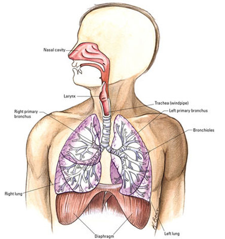
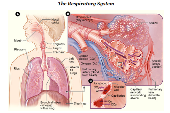
Nose- The nose is the entrance to the respiratory system and as air enters the nasal cavity it travels to and from the lungs. The thin layer of tissue —mucous membrane— and the fine hairs that line the nose help warm, moisten, and filter the air as it flows toward the lungs. The interior of the nose, or nasal cavity, is separated by a midline partition, known as the nasal septum. The paranasal sinuses are air-containing cavities that are lined with mucous membrane in the bones around the nose. The paranasal sinuses produce secretions that drain into, and lubricate, the nasal cavity.
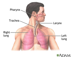
Pharynx- From the nasal cavity, air passes into the pharynx. The pharynx, also called the throat, serves as a passageway for food and air. Contained within the pharynx are the adenoids and the tonsils. Both of these pairs of organs are masses of lymphatic tissue that help to filter bacteria—especially the organisms that invade the body through the nose and mouth.
Nasopharynx- Located above the soft palate, the nasopharynx communicates with the nasal cavity and provides a passageway for air during breathing. The eustachian tubes, which connect the pharynx with the middle ears, open through the walls of the nasopharynx.
Larynx- The pharynx opens into the larynx and the esophagus. The larynx—also called the voice box—forms part of the passageway for air to the lungs. In addition to warming, humidifying, and filtering incoming air, the larynx contains the vocal cords.
Lower Respiratory System:
Trachea- The trachea, or windpipe, is a cylindrical tube in the neck. The trachea extends from the larynx to divide into two large air passages called the bronchial tubes. The trachea begins right below larynx, then it divides into two smaller tubes known as bronchi; each lung has a bronchus. Each time you inhale, the trachea slightly lengthens and widens, and it goes back to its normal size whenever you exhale.
Bronchi- The bronchi allow the passage of air to the lungs. The trachea is made of c-shaped ringed cartilage that divides into the right and left bronchus. The right main bronchus is shorter and wider than the left main bronchus. The right bronchus is subdivided into three lobar bronchi, while the left one is divided into two lobar bronchi.
Lungs- The lungs are spongy, air-filled organs located on both sides of the chest cavity. The left lung is divided into a superior and inferior lobe, and the right lung is subdivided into a superior, middle, and inferior lobe. Pleura, a thin layer of tissue, line the lungs to allow the lungs to expand and contract with ease. Respiration is the primary function of the lungs, which includes the transfer of oxygen found in the atmosphere into the blood stream and the release of carbon dioxide into the air.
Alveoli- The average adult has about 600 million alveoli, which are tiny grape-like sacs at the end of the respiratory tree. The exchange of oxygen and carbon dioxide gases occurs at the alveolar level. Although effort is required to inflate the alveoli (similar to blowing up a balloon), minimal effort is needed to deflate the alveoli (similar to the deflating of a balloon).
Diaphragm- The diaphragm is a muscular structure located between the thoracic and abdominal cavity. Contraction of the diaphragm causes the chest or thorax cavity to expand, which occurs during inhalation. During exhalation, the release of the diaphragm causes the chest or thoracic cavity to contract.
Nose- The nose is the entrance to the respiratory system and as air enters the nasal cavity it travels to and from the lungs. The thin layer of tissue —mucous membrane— and the fine hairs that line the nose help warm, moisten, and filter the air as it flows toward the lungs. The interior of the nose, or nasal cavity, is separated by a midline partition, known as the nasal septum. The paranasal sinuses are air-containing cavities that are lined with mucous membrane in the bones around the nose. The paranasal sinuses produce secretions that drain into, and lubricate, the nasal cavity.
Pharynx- From the nasal cavity, air passes into the pharynx. The pharynx, also called the throat, serves as a passageway for food and air. Contained within the pharynx are the adenoids and the tonsils. Both of these pairs of organs are masses of lymphatic tissue that help to filter bacteria—especially the organisms that invade the body through the nose and mouth.
Nasopharynx- Located above the soft palate, the nasopharynx communicates with the nasal cavity and provides a passageway for air during breathing. The eustachian tubes, which connect the pharynx with the middle ears, open through the walls of the nasopharynx.
Larynx- The pharynx opens into the larynx and the esophagus. The larynx—also called the voice box—forms part of the passageway for air to the lungs. In addition to warming, humidifying, and filtering incoming air, the larynx contains the vocal cords.
Lower Respiratory System:
Trachea- The trachea, or windpipe, is a cylindrical tube in the neck. The trachea extends from the larynx to divide into two large air passages called the bronchial tubes. The trachea begins right below larynx, then it divides into two smaller tubes known as bronchi; each lung has a bronchus. Each time you inhale, the trachea slightly lengthens and widens, and it goes back to its normal size whenever you exhale.
Bronchi- The bronchi allow the passage of air to the lungs. The trachea is made of c-shaped ringed cartilage that divides into the right and left bronchus. The right main bronchus is shorter and wider than the left main bronchus. The right bronchus is subdivided into three lobar bronchi, while the left one is divided into two lobar bronchi.
Lungs- The lungs are spongy, air-filled organs located on both sides of the chest cavity. The left lung is divided into a superior and inferior lobe, and the right lung is subdivided into a superior, middle, and inferior lobe. Pleura, a thin layer of tissue, line the lungs to allow the lungs to expand and contract with ease. Respiration is the primary function of the lungs, which includes the transfer of oxygen found in the atmosphere into the blood stream and the release of carbon dioxide into the air.
Alveoli- The average adult has about 600 million alveoli, which are tiny grape-like sacs at the end of the respiratory tree. The exchange of oxygen and carbon dioxide gases occurs at the alveolar level. Although effort is required to inflate the alveoli (similar to blowing up a balloon), minimal effort is needed to deflate the alveoli (similar to the deflating of a balloon).
Diaphragm- The diaphragm is a muscular structure located between the thoracic and abdominal cavity. Contraction of the diaphragm causes the chest or thorax cavity to expand, which occurs during inhalation. During exhalation, the release of the diaphragm causes the chest or thoracic cavity to contract.
Physiology of the Respiratory system
Pulmonary Ventilation- The process of moving air into and out of the lungs to facilitate gas exchange. The respiratory system uses a negative pressure system and the contraction of muscles to achieve pulmonary ventilation. The negative pressure gradient is established between the alveoli and the external atmosphere.The pleural membrane seals the lungs and maintains a pressure slightly below the pressure of the atmosphere while the lungs are at rest. The result is air follows the pressure gradient and passively fills the lungs at rest. As the lungs expand with air, the pressure within the lungs increases until it matches the atmospheric pressure. At this point, more air can be inhaled through the contraction of the diaphragm and the external intercostal muscles. This reduces the pressure of the lungs below the pressure of the atmosphere by increasing the volume of the thoracic cavity.
To exhale, the diaphragm and the external intercostal muscles relax while the internal intercostal muscles contract to decrease the volume of the thorax and increase the pressure in the thoracic cavity. This reverses the pressure gradient and results in the exhalation of air until the pressure inside and outside of the lungs reaches equilibrium. At this point, the lungs recoil back to their resting volume, which restores the negative pressure gradient present during inhalation.
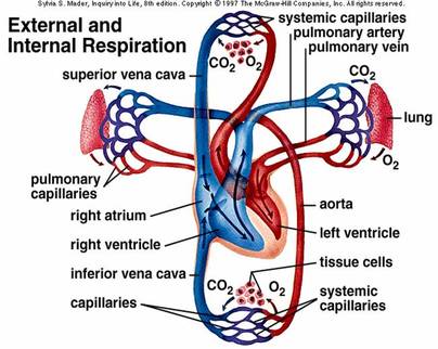
External Respiration- The exchange of gases between the air filling the alveoli and the blood in the capillaries that surround the walls of the alveoli. The air that enters the lungs from the atmosphere has a higher partial pressure of oxygen and a lower partial pressure of carbon dioxide than the blood in the capillaries. The difference in partial pressure causes the gases to diffuse passively along their pressure gradients from a high to low concentration through the lining of the alveoli. The net result of external respiration is the movement of oxygen from the air into the blood and the transportation of carbon dioxide from the blood into the air. The oxygen can then be transported to the body tissues while carbon dioxide is released to the atmosphere during exhalation.
Internal Respiration- The exchange of gases between the blood in capillaries and the tissues of the body. Capillary blood has a higher partial pressure of oxygen and a lower partial pressure of carbon dioxide than the tissues through which it passes. The partial pressure difference leads to the diffusion of gases along their pressure gradients from high to low pressure through the lining of the capillaries. The net result of internal respiration is the diffusion of oxygen into the tissues and the diffusion of carbon dioxide into the blood.
Transportation of Gases- The two major respiratory gases, oxygen and carbon dioxide, are transported through the body in the blood. A majority of the gases transported through the blood are bonded to transport molecules. Hemoglobin carries nearly 99% of the oxygen in the blood and is found in the red blood cells. While hemoglobin transports oxygen, carbon dioxide mainly transported in the plasma as bicarbonate ions.
Information from this page was gathered from: http://www.innerbody.com/anatomy/respiratory
To exhale, the diaphragm and the external intercostal muscles relax while the internal intercostal muscles contract to decrease the volume of the thorax and increase the pressure in the thoracic cavity. This reverses the pressure gradient and results in the exhalation of air until the pressure inside and outside of the lungs reaches equilibrium. At this point, the lungs recoil back to their resting volume, which restores the negative pressure gradient present during inhalation.
External Respiration- The exchange of gases between the air filling the alveoli and the blood in the capillaries that surround the walls of the alveoli. The air that enters the lungs from the atmosphere has a higher partial pressure of oxygen and a lower partial pressure of carbon dioxide than the blood in the capillaries. The difference in partial pressure causes the gases to diffuse passively along their pressure gradients from a high to low concentration through the lining of the alveoli. The net result of external respiration is the movement of oxygen from the air into the blood and the transportation of carbon dioxide from the blood into the air. The oxygen can then be transported to the body tissues while carbon dioxide is released to the atmosphere during exhalation.
Internal Respiration- The exchange of gases between the blood in capillaries and the tissues of the body. Capillary blood has a higher partial pressure of oxygen and a lower partial pressure of carbon dioxide than the tissues through which it passes. The partial pressure difference leads to the diffusion of gases along their pressure gradients from high to low pressure through the lining of the capillaries. The net result of internal respiration is the diffusion of oxygen into the tissues and the diffusion of carbon dioxide into the blood.
Transportation of Gases- The two major respiratory gases, oxygen and carbon dioxide, are transported through the body in the blood. A majority of the gases transported through the blood are bonded to transport molecules. Hemoglobin carries nearly 99% of the oxygen in the blood and is found in the red blood cells. While hemoglobin transports oxygen, carbon dioxide mainly transported in the plasma as bicarbonate ions.
Information from this page was gathered from: http://www.innerbody.com/anatomy/respiratory
