Digestive System
Overview of the digestive system
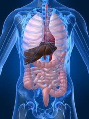
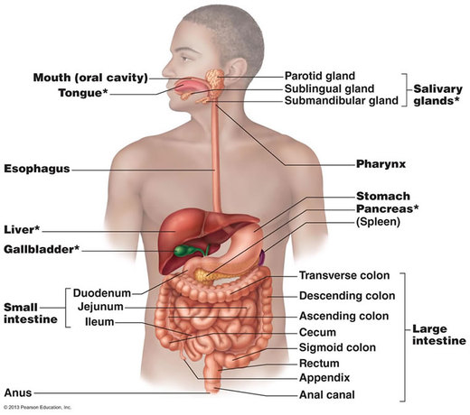
The digestive system is a group of organs that function together to convert food into energy and nutrients for the body. Food passes through a tube in the body called the alimentary canal, also known as the gastrointestinal tract (GI tract). The alimentary canal is composed of the oral cavity, pharynx, esophagus, stomach, small intestines, and large intestines. Accessory organs of the digestive system include the teeth, tongue, salivary glands, liver, gallbladder, and pancreas. To provide energy and nutrients to the body, six major functions take place in the digestive system: ingestion, secretion, mixing and movement, digestion, absorption, and excretion.
Anatomy of the Digestive system
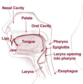
Digestive System of the Head and Neck: The initial digestive process occurs in the head and the neck where structures are responsible for the ingestion, chewing, and swallowing of food. These structures include the parts of the mouth, the salivary glands, and the epiglottis.
Mouth- Known as the oral cavity, the mouth allows air and food to enter into the body and begins the initial digestion process.
Teeth- Teeth are used to masticate (chew) food into tiny pieces. Classified into four groups, teeth are composed of incisors, canines, premolars, and molars.
Tongue- The tongue is made up of muscles covered by mucous membranes. There are small nodules, called papillae, on the top surface of the tongue, which give it its rough texture. Between the papillae at the sides and base of the tongue are small sensory organs, called taste buds, which enable the sensation of flavor. The muscle fibers of the tongue are heavily supplied with nerves, which allows food in the mouth to be manipulated and placed between the teeth for chewing. The tongue also coordinates its movements to aid in swallowing.
Salivary Glands- Surrounding the mouth are 3 sets of salivary glands. Saliva helps to moisten food and begins the digestion of carbohydrates. The body also uses saliva to lubricate food as it passes through the mouth, pharynx, and esophagus.
Soft Palate- The soft palate forms a muscular arch that extends back and down the posterior portion of the mouth toward the uvula. During swallowing, muscles draw the soft palate and the uvula upward. This allows the opening between the nasal cavity and the pharynx to close and prevents food from entering the nasal cavity.
Epiglottis- Composed of elastic cartilage, the epiglottis is a leaf-shaped structure at the superior portion of the larynx, while relaxed, it projects into the pharynx. It acts as a switch between the larynx and the esophagus to permit air to enter the pathway to the lungs and food to pass into the gastrointestinal tract.
Digestive System of the Upper Torso: The upper torso region of the digestive system is also known as the upper gastrointestinal tract. It includes several of the most important organs of the digestive system such as the esophagus, stomach, duodenum, liver, and gallbladder. These organs are responsible for the significant breakdown of food and the transportation of partially digested food further through the alimentary canal. After reaching the stomach, food is stored and digested with acid and digestive enzymes to form a liquid called chyme. Next, the chyme passes into the first segment of the small intestine called the duodenum. Bile from the liver and gallbladder along with enzymes from the pancreas are added to chyme in the duodenum to further digest it into small nutrients that can be absorbed by the intestines.
Esophagus- A long, thin, muscular tube that connects the pharynx to the stomach. A vital portion of the alimentary canal or GI tract, the esophagus assists in channeling food and liquids into the pharynx and to the stomach.
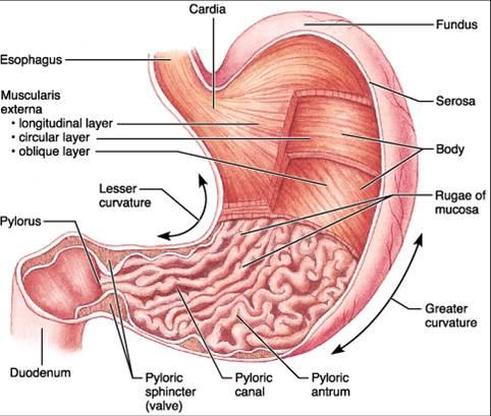
Stomach- The stomach is the main storage facility of food in the body. The stomach secretes a mixture of acid, mucus, and digestive enzymes to assist in the digestion of food while it is being stored. The inner portion of the stomach is comprised of wrinkles which are called rugae. The rugae allow the stomach to stretch, grip, and move food during digestion. The stomach is divided in to four regions, the cardia, the body, the fundus, and the pylorus. The concave medial border of the stomach is called the lesser curvature and the convex lateral border is called the greater curvature.
Mouth- Known as the oral cavity, the mouth allows air and food to enter into the body and begins the initial digestion process.
Teeth- Teeth are used to masticate (chew) food into tiny pieces. Classified into four groups, teeth are composed of incisors, canines, premolars, and molars.
Tongue- The tongue is made up of muscles covered by mucous membranes. There are small nodules, called papillae, on the top surface of the tongue, which give it its rough texture. Between the papillae at the sides and base of the tongue are small sensory organs, called taste buds, which enable the sensation of flavor. The muscle fibers of the tongue are heavily supplied with nerves, which allows food in the mouth to be manipulated and placed between the teeth for chewing. The tongue also coordinates its movements to aid in swallowing.
Salivary Glands- Surrounding the mouth are 3 sets of salivary glands. Saliva helps to moisten food and begins the digestion of carbohydrates. The body also uses saliva to lubricate food as it passes through the mouth, pharynx, and esophagus.
- Parotid Glands- The largest salivary glands located on either side of the jaw, anterior to the ears. The parotid glands secrete saliva into the posterior of the mouth.
- Submandibular Glands- Anterior and slightly inferior to the parotid glands, the submandibular glands rest on the inner side of the jaw and secrete saliva into the medial portion of the mouth.
- Sublingual glands- Inferior to the tongue, the sublingual glands secrete saliva into the anterior portion of the mouth.
Soft Palate- The soft palate forms a muscular arch that extends back and down the posterior portion of the mouth toward the uvula. During swallowing, muscles draw the soft palate and the uvula upward. This allows the opening between the nasal cavity and the pharynx to close and prevents food from entering the nasal cavity.
Epiglottis- Composed of elastic cartilage, the epiglottis is a leaf-shaped structure at the superior portion of the larynx, while relaxed, it projects into the pharynx. It acts as a switch between the larynx and the esophagus to permit air to enter the pathway to the lungs and food to pass into the gastrointestinal tract.
Digestive System of the Upper Torso: The upper torso region of the digestive system is also known as the upper gastrointestinal tract. It includes several of the most important organs of the digestive system such as the esophagus, stomach, duodenum, liver, and gallbladder. These organs are responsible for the significant breakdown of food and the transportation of partially digested food further through the alimentary canal. After reaching the stomach, food is stored and digested with acid and digestive enzymes to form a liquid called chyme. Next, the chyme passes into the first segment of the small intestine called the duodenum. Bile from the liver and gallbladder along with enzymes from the pancreas are added to chyme in the duodenum to further digest it into small nutrients that can be absorbed by the intestines.
Esophagus- A long, thin, muscular tube that connects the pharynx to the stomach. A vital portion of the alimentary canal or GI tract, the esophagus assists in channeling food and liquids into the pharynx and to the stomach.
Stomach- The stomach is the main storage facility of food in the body. The stomach secretes a mixture of acid, mucus, and digestive enzymes to assist in the digestion of food while it is being stored. The inner portion of the stomach is comprised of wrinkles which are called rugae. The rugae allow the stomach to stretch, grip, and move food during digestion. The stomach is divided in to four regions, the cardia, the body, the fundus, and the pylorus. The concave medial border of the stomach is called the lesser curvature and the convex lateral border is called the greater curvature.
- Cardia: The point at which the esophagus connects to the stomach. The cardia is a narrow tube structure that and opens into the wider region of the stomach. In the cardia is the cardiac sphincter also known as the lower esophageal sphincter, a circular band of muscle that closes the end portion of the esophagus and prevents the stomach contents from flowing backward.
- Body: The body receives everything from the cardia, and forms the central and largest region of the stomach.
- Fundus: Superior to the body, the fundus forms a dome shape
- Pylorus: Inferior to the body, the pylorus forms a funnel shape. The pylorus connects the stomach to the duodenum and houses the pyloric sphincter. This sphincter controls the flow of partially digested chyme out of the stomach and into the duodenum.
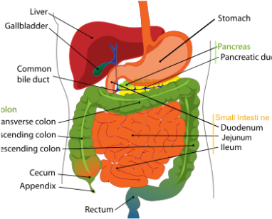
Liver- The liver is the body's second largest organ, it performs essential functions related to digestion, metabolism, and storage of nutrients in the body. The liver has multiple bile ducts that transport bile and it is drained into microscopic canals known as bile canaliculi. The bile canaliculi join together into many larger bile ducts found throughout the liver.
Gallbladder- Located inferior and posterior to the liver, the gallbladder holds bile from the liver until it is needed to digest fatty foods in the duodenum from the small intestine. One end of the gallbladder narrows into the cystic bile duct. Mucosa lines the inner layer of the gallbladder and bile slowly concentrates through the absorption of water through the walls of the gallbladder. The contraction of smooth muscle forces bile out of the gallbladder and into the cystic duct. From the cystic duct, bile enters the common bile duct and flows into the ampulla of Vater, where the bile ducts merge with the pancreatic duct. Bile then flows into the duodenum.
Pancreas- A glandular organ, the pancreas serves as a digestive exocrine gland that excretes enzymes to break down the proteins, lipids, carbohydrates, and nucleic acids in food. The pancreatic duct carries the digestive enzymes produced by endocrine cells to the duodenum.
Small Intestine- The small intestine absorbs nearly 90% of the nutrients we receive from our food. The small intestine is divided into three regions, the duodenum, the jejunum, and the ileum. A membrane called mesentery surrounds the small intestine and anchors it in place. The small intestine is composed of four layers of tissue, (from deep to superficial) submucosa, mucosa, muscularis layer, and serosa. The three regions of the small intestine are explained as follows:
- Heptatic ducts: Carry bile from the left and right lobes of the liver. The hepatic ducts connect to form the common hepatic duct, which drains the bile away from the liver. The common hepatic duct joins with the cystic duct from the gallbladder and forms the common bile duct.
- Common bile duct: The common bile duct carries bile to the duodenum of the small intestine. Most of the bile produced by the liver is pushed back up the cystic duct by peristalsis to arrive in the gallbladder for storage, until it is needed for digestion.
Gallbladder- Located inferior and posterior to the liver, the gallbladder holds bile from the liver until it is needed to digest fatty foods in the duodenum from the small intestine. One end of the gallbladder narrows into the cystic bile duct. Mucosa lines the inner layer of the gallbladder and bile slowly concentrates through the absorption of water through the walls of the gallbladder. The contraction of smooth muscle forces bile out of the gallbladder and into the cystic duct. From the cystic duct, bile enters the common bile duct and flows into the ampulla of Vater, where the bile ducts merge with the pancreatic duct. Bile then flows into the duodenum.
Pancreas- A glandular organ, the pancreas serves as a digestive exocrine gland that excretes enzymes to break down the proteins, lipids, carbohydrates, and nucleic acids in food. The pancreatic duct carries the digestive enzymes produced by endocrine cells to the duodenum.
Small Intestine- The small intestine absorbs nearly 90% of the nutrients we receive from our food. The small intestine is divided into three regions, the duodenum, the jejunum, and the ileum. A membrane called mesentery surrounds the small intestine and anchors it in place. The small intestine is composed of four layers of tissue, (from deep to superficial) submucosa, mucosa, muscularis layer, and serosa. The three regions of the small intestine are explained as follows:
- Duodenum: The first section of the intestine, the duodenum connects to the pyloric sphincter of the stomach. Partially digested food (also known as chyme) from the stomach, mixes with bile from the liver and pancreatic juice to complete the duodenum digestive process.
- Jejunum: The middle section of the small intestine, the jejunum serves as the primary absorption site of nutrients.
- Ileum: The final section of the small intestine, the ileum empties into the large intestine through the ileocecal sphincter.
Digestive System of the Lower Torso:
Cecum- The pouch where the large intestine begins, it is where the ileum opens from one side and continues with the colon.
Ascending Colon- Carries feces from the cecum to the transverse colon. The intestinal wall absorbs water, nutrients, and vitamins from the feces and deposits the materials into the bloodstream.
Transverse Colon- The largest part of the colon, it attaches the ascending colon to the descending colon by crossing the abdominal cavity.
Descending Colon- Transports feces from the transverse colon and stores it in the descending colon until it is ready to be eliminated from the body. The walls of the descending colon absorb water as well as the remaining nutrients and vitamins from the feces.
Sigmoid Colon- The final segment of the colon, the sigmoid colon transports fecal matter from the descending colon to the rectum and anus.
Rectum- A muscular tube that forms the lowest portion of the large intestine and connects it to the anus.
Anus- The anus is a canal at the end of the digestive tract where feces is expelled.
Information from this page was gathered from: http://www.innerbody.com/image/endoov.html
